
Pain in front of the ear, joint noises, difficulty opening the mouth, or a feeling that the jaw „locks up” are among the most common reasons patients seek a temporomandibular joint (TMJ) specialist.
And yet, one of the most frequently asked questions is:
„If I've already had an X-ray or CT scan, why do I need an MRI?”
The answer is simple: because the most important structures involved in many TMJ dysfunctions It's not bones, you Articular cartilage, ligaments, joint capsule, synovial fluid, and soft tissues, and these can only be fully evaluated by magnetic resonance imaging (MRI).
Today, MRI represents the gold standard for examining the soft tissues of the temporomandibular joint and plays an essential role in establishing a correct diagnosis and personalized treatment.
What is the temporomandibular joint (TMJ)?
The temporomandibular joint is one of the most complex joints in the body.
It allows:
- mouth opening;
- closing the mouth;
- chewing;
- speech;
- swallowing;
- sideways and forward movements of the mandible.
Unlike many other joints, the TMJ contains a Articular fibrocartilaginous disc which functions as a shock absorber between the mandibular condyle and the temporal bone.
When this disc no longer functions correctly or changes its position, symptoms such as the following may appear:
- starttours;
- Precisely;
- blockage;
- pain;
- limited mouth opening;
- The sensation of the jaw „slipping out of place.”.
All these changes can be analyzed in detail by MRI.
Why aren't X-rays or CT scans sufficient?
This is probably the most important question. X-rays and CBCT are excellent for evaluating bone.
They can highlight:
- fractures;
- erosion;
- osteophytes;
- bone remodeling;
- Ankylosis;
- advanced degenerative changes.
Give him I can't show:
- articular disc;
- ligaments;
- capsule;
- synovial fluid;
- synovitis;
- bone marrow edema;
- early inflammatory changes.
Cu alte cuvinte…
You can have a perfectly normal CT scan and still have temporomandibular joint dysfunction.
This is one of the reasons why numerous patients receive the answer for years:
„You have nothing.”
Although their symptoms are real.
What can an MRI of the TMJ see?
MRI provides information that cannot be obtained through other imaging methods.
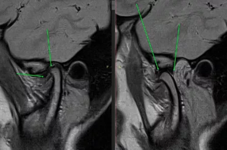
Among the most important structures evaluated are:
- Joint pain
This is the main reason an ATM MRI is performed. It can assess:
- disk position;
- its form;
- thickness;
- potential deformations;
- perforations;
- previous movement;
- medial displacement;
- lateral displacement;
- reduction during mouth opening.
- Ligamentsand capsule articulate
MRI allows for the evaluation of ligamentous apparatus stability and capsular changes that can occur following trauma or chronic overuse.
- Joint inflammation
A major advantage of MRI is its ability to identify inflammation even before bone begins to deteriorate.
Can be observed:
- sinovita;
- Joint fluid buildup;
- thickening of the synovial membrane;
- Periarticular inflammatory reaction.
These changes can explain the pain even when the X-ray is normal.
- Mandibular condyle
Although CBCT remains superior for bone cortical detail, MRI provides extremely valuable information about:
- bone marrow;
- bone marrow biopsy;
- necrosis;
- early degenerative changes;
- vasculature.
- Musclesandtissues neighboring
In certain situations, the following can also be evaluated:
- Masticatory musculature;
- tendon of the lateral pterygoid muscle;
- inflammatory collections;
- tumors;
- post-traumatic changes.
Why is the MRI done with the mouth closed and with the mouth open?
Many patients are surprised when they learn that the exam does not involve a single position. In reality, a proper assessment requires examining the joint in Occlusion (closed mouth), as well as maximum comfortable opening.
Why? Because the articular disc is a mobile structure.
A disc that appears to be displaced with its mouth closed may return completely to its position during opening. This is called: Reduced disk displacement.
Instead, if the disc remains displaced even during opening, we are talking about: displacement without reduction, a situation frequently associated with limited mouth opening and joint locking.
This is why modern MRI protocols include dedicated sequences in both positions, in oblique planes adapted to the TMJ anatomy.
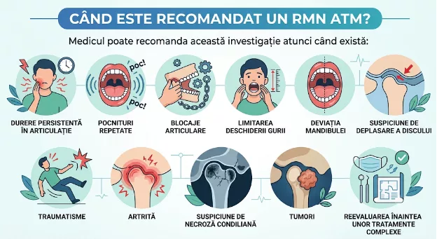
When is an ATM MRI recommended?
The doctor may recommend this investigation when there is:
- persistent joint pain;
- repeated strokes;
- joint block;
- limited mouth opening;
- jaw deviation;
- Suspicion of disc displacement;
- trauma;
- arthritis;
- suspicion of condylar necrosis;
- tumors;
- reevaluation before complex treatments.
What do studies say?
Numerous studies published in international literature have shown that MRI represents method imagery of reference for evaluation structures I want an ATM. This allows for simultaneous analysis of:
- article hinge;
- ligaments;
- synovial fluid;
- synovial membrane;
- bone marrow;
- inflammatory changes;
- the relationship between the condyle and disc during movement.
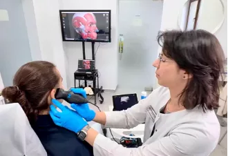
Additionally, current guidelines recommend correlating MRI findings with the clinical examination, as treatment should be established based on both symptoms and imaging changes. Not every visible change on MRI requires intervention, and some individuals may present with disc displacement without pain or functional limitation.
If the MRI is normal, why does it hurt?
This is one of the most important questions. Pain in the TMJ region does not always originate from the joint. It can originate from:
- Masticatory musculature;
- neck musculature;
- cervical fascia;
- myofascial trigger points;
- muscle overuse;
- bruxism;
- postural disorders;
- neurological conditions;
- central sensitization.
This is why interpreting an MRI in isolation, without examining the patient, can lead to incomplete conclusions.
7 myths about TMJ MRI
False.
X-rays cannot evaluate the articular disc and other soft tissues.
False.
The majority of cases initially benefit from conservative treatments, adapted to the cause and severity of symptoms.
No.
The investigation is completely non-invasive and painless.
No.
The quality of the investigation depends on the protocol used, the MRI device, and the radiologist's experience in temporomandibular joint imaging.
Not always.
The goal of treatment is pain reduction and restoration of function, not necessarily perfect anatomical repositioning of the disc.
It depends on what we're looking for.
CT is superior for bone. MRI is superior for the articular disc and soft tissues.
The two investigations are complementary.
No.
He represents just one piece of the puzzle. The correct diagnosis emerges when the images are integrated with the patient's history, clinical examination, functional assessment, and, when necessary, other imaging investigations.
Jaw pain, joint noises, and limited jaw movement should not be considered „normal” or ignored. In many situations, the cause of symptoms cannot be identified through a simple X-ray or CBCT scan, as it involves the soft tissues of the temporomandibular joint.
The ATM MRI is today the reference investigation for evaluating the articular disc, ligaments, capsule, and inflammatory processes, providing essential information for an accurate diagnosis and for choosing the most appropriate treatment.
However, the most valuable investigation is not the one that produces the most images, but the one that is interpreted in the patient's clinical context. An MRI should be correlated with symptoms, functional examination, muscle analysis, occlusal evaluation, and, when necessary, postural analysis. Only this integrated approach allows for the identification of the real cause of the pain and the construction of a personalized therapeutic plan aimed at functional recovery and improved quality of life.
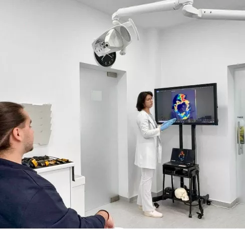
The Innovation Medical Center, We believe that correct treatment always begins with a correct diagnosis. That is why the evaluation of patients with temporomandibular joint dysfunction is not based solely on imaging, but on integrating MRI results with a clinical examination, functional analysis, muscle evaluation, occlusal evaluation, and, when necessary, postural analysis. This multidisciplinary approach allows us to identify the real cause of symptoms and develop a personalized therapeutic plan, based on scientific evidence and tailored to each patient.
Schedule a comprehensive evaluation
📍 București – www.innovationmedical.ro
📞 0753 666 111
Autor: Dr. Anca Adam, Medic Specialist in Medicina Fizica si de Reabilitare, compartiment recuperare neurologică și disfuncții ATM/TMJ, Expert Acupunctură Laser & Competenta Medicina Traditionala Chineza (Tai Yuan China), Termografie Medicală (Barcelona, Spania) – LSO (Laser Safety Officer) AALZ Aachen Germania.




















 Romanian
  English