
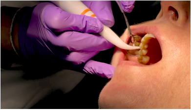
Periodontal disease is one of the most common chronic inflammatory conditions and is the leading cause of tooth loss in adults. It progressively affects the gums, periodontal ligament, and alveolar bone – the structures that support the teeth. In the absence of adequate treatment, inflammation and infection cause the destruction of these tissues, tooth mobility, and ultimately, tooth loss.
One of the biggest challenges is that the disease often progresses without pain. Many patients seek treatment only when they notice persistent bleeding gums, gum recession, bad breath, or tooth mobility, even though the destruction of supporting tissues is already advanced.
In recent years, numerous studies have shown that oral health and general health are profoundly interconnected. Chronic inflammation associated with periodontal disease can influence the progression of systemic conditions such as cardiovascular disease, diabetes mellitus, respiratory diseases, certain pregnancy complications, and is associated with an increased risk of cognitive decline and the development of Alzheimer's disease.
Why isn't a visual examination alone sufficient?
Gum appearance provides only partial information. Bone destruction and attachment loss occur below the gum line, in areas that cannot be accurately assessed by clinical inspection alone.
Therefore, a comprehensive periodontal examination involves measuring each tooth at six different points, thus obtaining hundreds of values for the entire oral cavity.
These measurements allow for the determination of disease severity, selection of treatment, and monitoring of results over time.
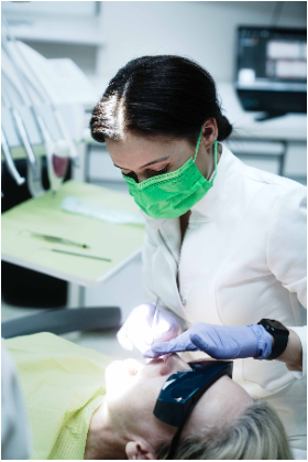
The Importance of Digital Periodontal Probing
In modern dentistry, periodontal examination can be performed with the help of a digital probe, which electronically records each measurement and reduces the variability associated with manual probing.
In addition to measuring periodontal pocket depth, the system allows for the evaluation of clinical attachment loss, recording bleeding on probing, assessing plaque index, tooth mobility, and furcation involvement. All of this information is stored digitally and can be objectively compared at each visit.
The result is not just a set of numbers, but a complete picture of the health of the periodontium.
What do the graphs show us?
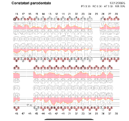
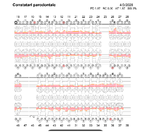
The graphs below illustrate the progression of the same patient before and after periodontal treatment.
Îbefore treatment
- numerous deep periodontal pockets.;
- gingival bleeding on probing;
- inflammation;
- increased risk of disease progression and tooth loss.
După treatment
- significant reduction in bag depth;
- reduced bleeding on catheterization;
- reducing gum inflammation;
- stabilization of supporting tissues;
- Improved long-term prognostic.
Why do differences of only 1-2 mm matter?
In periodontics, a single millimeter can change the diagnosis, prognosis, and even the therapeutic plan.
Therefore, measurements must be as reproducible and comparable as possible between visits. Digital systems reduce examination variability and allow for objective patient monitoring, providing the clinician with accurate information for each stage of treatment.
Monitoring is as important as treatment
Periodontal treatment does not end after scaling or initial therapy.
Periodontal disease is a chronic condition and requires regular monitoring. Digital comparison of measurements allows for rapid identification of stable areas and those where the disease continues to progress, before the patient notices obvious symptoms.
How do we interpret the depth of periodontal pockets?
The depth of the periodontal pocket represents the distance between the gingival margin and the deepest point a periodontal probe can penetrate. The greater this value, the more advanced the destruction of the tooth's supporting tissues.
In general, the interpretation is as follows:
1–3 mm – Situationțphysiologicallyă
These values are considered normal for most patients. The supporting tissues are stable, and the gums are healthy, with no inflammation or bleeding.
4 mm – Alarm signală
It can indicate the onset of a periodontal condition or the presence of persistent gum inflammation. At this stage, treatment is usually simpler, and the prognosis is very good if the patient follows the doctor's recommendations.
5–6 mm – Boală periodontală moderateă
The pockets become deep enough that bacteria can no longer be effectively removed by brushing and flossing alone. Professional periodontal treatment is needed to control the infection and prevent disease progression.
7 mm or more – Boală periodontală advancedă
These values typically indicate significant bone loss and periodontal attachment loss. The risk of tooth mobility and tooth loss increases significantly if treatment is not initiated in a timely manner.
Depth isn't the only thing that matters
A bag of 5 millimeters it doesn't always have the same meaning.
The doctor always interprets these values along with other clinical parameters:
- Bleeding on catheterization
- loss of clinical attachment
- presence of bacterial plaque;
- tooth mobility;
- furcation involvement in multi-rooted teeth;
- Radiographic evaluation of bone level.
That's why two people with the same pocket depth can have completely different diagnoses and treatment plans.
The objective of treatment is not just reducing the numbers
Many patients believe that treatment success means all pockets reach 2-3 mm. In reality, the goal is to achieve a parodonțiu stable, without inflammation, bleeding on probing, and without mobility. In certain situations, some residual pockets may remain deeper, but if they are stable and show no signs of inflammatory activity, the long-term prognosis can be very good.
Advertisementâthe polling | SignifiesțEye clinică guidelineă |
1–3 mm | Values commonly found in a healthy periodontium, in the absence of inflammation. |
≥4 mm | Requires careful clinical evaluation along with other periodontal parameters. |
≥5 millimeters | Suggests the existence of a periodontal pocket requiring treatment and monitoring. |
Higher values | It can indicate more severe involvement, but the severity of the disease is not determined solely by the pocket depth. |
Important: The depth of periodontal pockets alone does not establish the diagnosis. The dentist always interprets these values along with bleeding on probing, clinical attachment loss, bone level on radiographs, tooth mobility, furcation involvement, and other clinical parameters.
Conclusion
In modern dentistry, treatment no longer solely relies on clinical experience, but also on decisions based on concrete data. Digital technologies allow for the measurement, documentation, and comparison of every periodontal parameter over time, providing both the clinician and the patient with a clear picture of the disease's progression and treatment effectiveness.
What cannot be measured cannot be accurately tracked. And what is not monitored cannot be truly controlled.

Programă or consultationțI.e. șand discoveră How modern laser technologies can transform dental treatments!
📍 Innovation Medical Center
Bucharest
📞 0753 666 111
AuthoriDr. Nur Saadeddin– Medical dentist within Innovation Medical Center, Co-Founder Cluj Smile Society, Expert in Laser dentistry, Master of Science – Laser therapy in dentistry (AALZ Aachen & Sigmund Freud University Vienna), Laser Safety Officer (LSO) – AALZ Aachen, trainer in laser therapies, with dedicated activity in prosthetics, periodontology, minimally invasive approaches, and digital dentistry.
Dr. Thomas Mendel– Medical Director and Dentist at Innovation Medical Center, Co-founder Cluj Smile Society, Laser Dentistry Expert, Master of Science – Laser therapy in dentistry (AALZ Aachen & Sigmund Freud University Vienna), Laser Safety Officer (LSO) – AALZ Aachen, trainer in laser dentistry, with dedicated activity in endodontics, periodontics, minimally invasive approaches, and digital dentistry.




















 Romanian
  English