

Imagine your jaw as a door that opens and closes 1,500–2,000 times a day. But it’s not a simple door—it’s a delicate construction between the temporal bone and the mandible, featuring a cartilaginous disc, controlled by muscles, nerves, ligaments, and microscopic choreography.
This joint is named TMJ – temporomandibular joint, and her „pivot” is called condiments.
If you have ear pain, jaw locking, popping, migraines, dizziness, neck pain, or a feeling that your jaw is „slipping,” chances are high that condylar movement to be affected.
In this article, I will explain in simple terms why is real-time 3D condylar movement evaluation essential and how this modern investigation changes the entire way we treat TMJ dysfunctions at Innovation Medical Center.
In 2026, at Innovation Medical Center, with digital technology at the 2026 level, you receive minimally invasive diagnosis and ATM therapy, plus complete dental treatments through laser dentistry, with fast and precise results from an interdisciplinary team. Innovation Medical Center is an associated center and European partner of Sigmund Freud University Vienna and AALZ Germany, being affiliated with ISLD, which guarantees clinical practices aligned with international academic and scientific standards in laser dentistry and ATM therapy and Medical Thermography.
Mandibular condyle
Condyle is Round jaw hinge enters the temporal cavity (glenoid fossa). Together with the articular disc, it forms the TMJ.
If the jaw were a bicycle, the condyle would be the „front wheel” that needs to move perfectly for the entire vehicle to function correctly.
What are the normal movements of the condyle?
The TMJ is one of the most complex joints in the body. The condyle not only „opens and closes” but also performs:
✔ 1. Rotation (first half of mouth opening)
The condyle rotates in place, like a cog on an axle.
✔ 2. Translation (second half of mouth opening)
The condyle slides forward and downward on a bony ramp called articular eminent.
✔ 3. Lateral movements (mastication)
One condyle rotates, the other translates — a perfectly coordinated movement.
✔ 4. Retraction and protrusion
The mandible moves forward and backward.
All of this must be symmetric, clean, and seamless to avoid pain and blockages.
⚠️ What happens when the condyle is no longer moving correctly?
Here they come TMJ pathologies, divided into:
Acut inflammation, swelling, pain, muscle spasms
Chronic → adaptations, fibrosis, displaced disc, joint wear, neuropathic phenomena
1. Blocked or limited movement
→ Limited opening, jaw deviates to the side.
2. Bearing with disc skip (click)
The disc is displaced, and the condyle jumps like a wheel on a curb.
3. Hypermobility
The condyle protrudes too much and dislocates easily.
4. TMJ Arthritis / Osteoarthritis
deformed condyle, irregular surfaces, pain on mastication.
How muscles and nerves suffer when the condyle moves abnormally
✔ The masseter and temporal muscles go into spasm → pain, tired face, dental wear.
✔ The lateral pterygoid pulls the discs → click, locking.
✔ Suprahyoid muscles modify swallowing → fatigue when speaking.
✔ Cervical chain (SCM, trapezius) becomes compensated → dizziness, headache, forward head posture.
✔ The auriculotemporal nerve can be irritated → pain in the ear, temple, and eye.
Pain can irradiate Spre
ear
eye
temple
cat
teeth
spate
That's why ATM it's not a local problem, a dysfunction Global, neuro-musculo-articular.
Why is an X-ray not enough?
Radiographs or CBCT show the shape of the condyle, but show movement.
It's like taking a picture of a door without checking if the hinges work.
Do you need dynamics, the „movie” of the joint, not just the pose.
3D evaluation of condyle movement – the key to correct treatment
This is where modern technologies come in:
✔ Axiography / Computerized Axiography
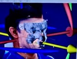
Devices such as Jaw Tracker SDI Matrix (https://sdimatrix.com/jaw-tracker/) measures in real time:
Condylar path
mandibular opening
rotation vs. translation
deviation, deflection
blockage
optimal functional position
The result?
A 3D model precise to the way your joint moves.
✔ Overlay of imaging investigations
Axiomatic data correlates with:
CBCT / TMJ radiography
RMN if needed
Real-time TMJ ultrasound (disc position, capsule inflammation)
thermographymuscle balance + compensatory chains
Postural assessment (head forward, shoulders rotating)
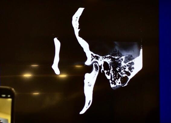
The functional position is The exact place where the jaw moves without pain and without muscular compensation.
If the jaw is „forced” into another position:
The condyle compresses nerve fibers.
disc can be pushed
muscle spasms
head posture changes
chronic pain
ATM treatment should only be done after the correct position has been established, otherwise the symptoms will return.
Comprehensive ATM Evaluation at Innovation Medical Center
The Innovation Medical Center, condylar movement is NOT analyzed in isolation.
🔹 3D Axiometry
Condylar movements are tracked millimeter by millimeter in all directions.
🔹 Overlay with imaging
CBCT + axiometrie + ultrasound → exact reconstruction of joint function.
🔹 Thermography
Identifying muscles in spasm and compensatory chains.
🔹 Postural and Neurological Analysis
The link between the TMJ, cervical spine, vertigo, and headaches.
🔹 Dental and occlusal evaluation
Dental contacts can force the jaw into pathological positions.
All of this data is integrated into specialized ATM assessment software, which calculates ideal functional position — the basis of multidisciplinary treatment.
Scientific studies
Recent studies support the importance of a comprehensive evaluation of the temporomandibular joint. First, the analysis conducted by Larheim TA et al. (2014) highlighting the role of CBCT in the morphological diagnosis of TMJ, as it is a reference method for evaluating the condyle, joint spaces, and degenerative bone changes. (Access: https://pmc.ncbi.nlm.nih.gov/articles/PMC4277441/)
Another important benchmark is the meta-analysis by Maranini B et al. (2022) care demonstrates the value of high-resolution ultrasonography in identifying articular disc displacement, offering a rapid and complementary method to traditional imaging investigations. (Access: https://pmc.ncbi.nlm.nih.gov/articles/PMC9251198/)
Also, the comparative study conducted by Yılmaz D et al. (2019) demonstrate that high-resolution ultrasonography can achieve accuracy similar to MRI for the diagnosis of internal derangements of the TMJ, including the detection of joint effusions, confirming its clinical value. https://pmc.ncbi.nlm.nih.gov/articles/PMC6747419/)
In addition, the prospective study by Abdel Razek AA et al. (2014) Demonstrate that ultrasound is useful for identifying anterior and lateral disc displacements in patients with temporomandibular joint pain and dysfunction. (Access: https://pubmed.ncbi.nlm.nih.gov/26191103/)
The study conducted by Osama A et al. (2023) Confirm that CBCT is a valuable tool in evaluating TMJ bone changes, joint spaces, and signs of osteoarthritis, being indispensable for structural analysis of the joint. (Access: https://pubmed.ncbi.nlm.nih.gov/40738007/)
In the end, his research Yu W et al. (2023) shows a clear correlation between changes in joint spaces visible on CBCT and disc displacement confirmed by MRI, emphasizing the importance of correlating imaging investigations with functional evaluation in establishing the correct mandibular position. (Access: https://pubmed.ncbi.nlm.nih.gov/38201353/)
❓ Frequently Asked Questions
Most commonly, the articular disc is displaced and the condyle jumps over it.
Now. Indicates muscle tension, bruxism, or condylar dysfunction.
Yes — the neuromuscular chain and the auriculotemporal nerve can cause vertigo and headaches.
Sound and pain can be „projected” into the ear through the nerves of the temporal area.
Only if the mandible is in a pathological position.
In over 90% of cases, yes—with muscle rebalancing, laser therapy, physical therapy, and functional mouthguards.
The Innovation Medical Center, each patient with temporomandibular dysfunction benefits from a comprehensive, multidisciplinary, and highly personalized evaluation. The integration of modern technologies — 3D axiography, dynamic TMJ ultrasound, thermography, computerized postural analysis, CBCT imaging, and functional dental assessment — allows for the precise identification of the actual cause of symptoms, not just the treatment of their effects. Our team works together to establish ideal functional position of the mandible essential for long-term stable treatments, whether it's joint pain, blockages, bruxism, migraines, dizziness, or postural imbalances. Our approach is based on functionality, precision, and interdisciplinary collaboration, so that each patient receives a comprehensive, safe, and effective treatment plan, built precisely for their needs. Innovation Medical Center thus becomes a modern benchmark in the diagnosis and treatment of TMD, offering real solutions and lasting results.
Autor: Dr. Anca Adam, Medic Specialist în Medicină Fizică și de Reabilitare, compartiment recuperare, medicină fizică și recuperare neurologică, disfuncții ATM/TMJ, Expert Acupunctură Laser, MTC & Termografie Medicală




















 Romanian
  English