
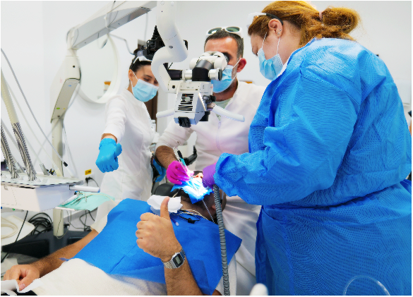
For many years, the operating microscope has become the gold standard instrument in endodontics, as it allows observation of details that are difficult or impossible to identify with the naked eye.
The root canal system is much more complex than it appears on an X-ray. Inside the roots, there are frequently extra canals, fine branches, and anatomical variations that can significantly influence the treatment outcome.
For this reason, all endodontic treatments in our practice are performed under a microscope and isolation.
You can't treat what you can't see
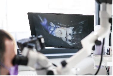
One of the most important advantages of the microscope is its ability to magnify the operating field and provide direct illumination inside the tooth.
This allows for the identification of situations that might go unnoticed without magnification.
Among these are:
- supplementary channel;
- calcified canals;
- perforations;
- fractured instruments;
- cracks;
- very fine anatomical details of the duct system.
In many cases, treatment success depends on identifying and treating these details.
Additional channels can explain the failure of some older treatments
One of the common causes of persistent infection is the existence of canals that were not previously identified and treated. These canals can harbor bacteria and infected tissue, even if the rest of the treatment was performed correctly. The microscope allows for their identification and treatment with much greater precision.
Calcified canals are one of the biggest challenges
As age advances or in response to trauma and inflammation, some channels can narrow or even partially close due to calcium deposits.
These situations are known as calcified canals. Their treatment requires patience, experience, and very good visual control. The microscope allows for the location and tracing of these canals, minimizing the risk of unnecessary removal of dental tissue. Fractured instruments do not automatically mean extraction. Contemporary endodontic instruments are very fine and work in extremely narrow spaces. In certain situations, they can fracture inside the canal. The microscope allows for an accurate assessment of the fragment's position and the selection of the most appropriate treatment strategy. Depending on the situation, the fragment can be removed, bypassed, or integrated into the treatment plan. The presence of a fractured instrument is not an indication for extraction.
Perforations can be diagnosed and treated more precisely
A perforation is an unwanted communication between the canal system and the tissues surrounding the root. Early diagnosis and correct treatment significantly influence the prognosis. The magnification provided by the microscope allows for precise localization of the perforation and control of repair procedures.
Microscope șin conservation țdental floss
Another important advantage is the possibility of working more conservatively. When visibility is increased, dental tissue removal can be strictly limited to the area necessary for treatment. Preserving as much healthy tooth structure as possible contributes to the tooth's strength and longevity.
Microscope not înlocuieșI can experienceța
The microscope does not treat the tooth. It is a tool that allows the dentist to see better and work with greater precision. The final outcome always depends on the diagnosis, experience, planning, and correct execution of the treatment. In modern endodontics, however, the microscope provides information that can make the difference between a predictable treatment and a failure.
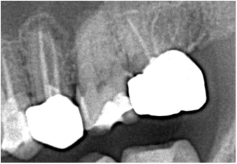
Radiograph 1 – Before retreatment - Previous endodontic treatment shows incomplete obturations, with poor delineation of the root canal system.
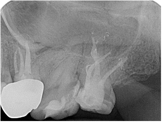
Radiograph 2 – After retreatment - Complete endodontic retreatment, with clear delineation of the main canals and highlighting of lateral and collateral canals.
Programă or consultationțI.e. șand discoveră How modern laser technologies can transform dental treatments!
📍 Innovation Medical Center
Bucharest
📞 0753 666 111
Authori:
Dr. Thomas Mendel– Medical Director and Dentist at Innovation Medical Center, Co-founder Cluj Smile Society, Laser Dentistry Expert, Master of Science – Laser therapy in dentistry (AALZ Aachen & Sigmund Freud University Vienna), Laser Safety Officer (LSO) – AALZ Aachen, trainer in laser dentistry, with dedicated activity in endodontics, periodontics, minimally invasive approaches, and digital dentistry.
Dr. Nur Saadeddin– Medical dentist within Innovation Medical Center, Co-Founder of Cluj Smile Society, Laser Dentistry Expert, Master of Science – Laser therapy in dentistry (AALZ Aachen & Sigmund Freud University Vienna), Laser Safety Officer (LSO) – AALZ Aachen, trainer in laser therapies, with dedicated activity in prosthetics, periodontology, minimally invasive approaches, and digital dentistry.




















 Romanian
  English